







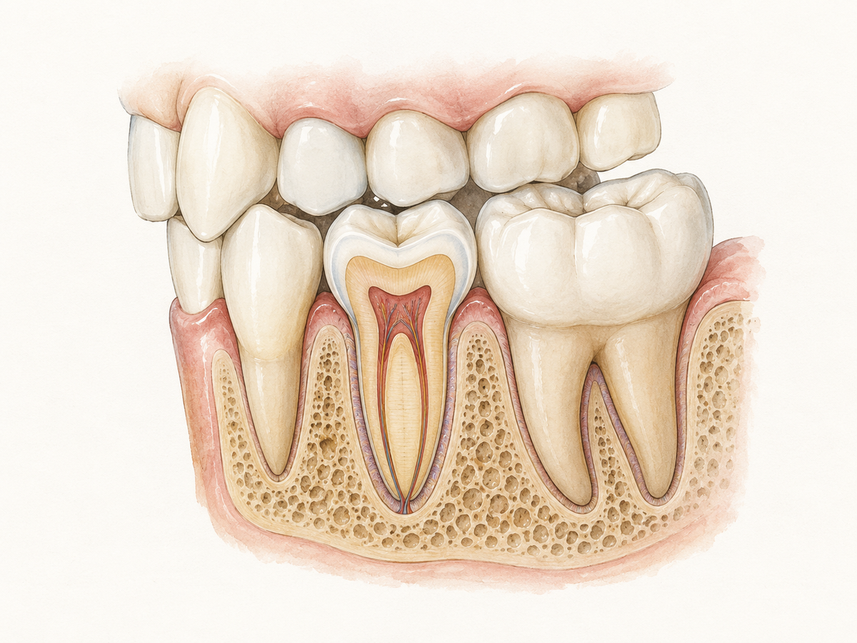
Premolars are the permanent in-between teeth that sit between your canines, the pointed eye teeth, and your molars, the wide grinding teeth at the back. Their main job is to help tear and crush food, but they also keep your bite aligned and support the shape of your cheeks. The truth is, they are not spare teeth. Losing one can quietly allow the teeth next to it to shift.
If a premolar feels loose, painful, or broken, that is a reason to see a dentist, not something to wait out.
What Premolars Actually Do
Premolars are often called transitional teeth because they bridge the gap between front-tooth and back-tooth functions. In everyday terms, they are the in-between teeth.
Their main job is to help the pointed canines tear food and to assist the molars with grinding. This happens during mastication, which simply means chewing. Because premolars handle both roles, they are not purely tearers or purely grinders. They do a bit of both.
Premolars do more than help you eat. They help upper and lower teeth meet correctly, which dentists call occlusion, or bite alignment. They also support the corners of your mouth and the shape of your cheeks. According to a dental anatomy reference on premolar function, premolars assist the canines in tearing food, supplement the grinding of the molars, and help maintain the vertical height of the bite.
That support matters. When one premolar goes missing, neighboring teeth can start to lean or drift into the open space. The change is often quiet at first, but over time it can affect how your whole bite feels.
Premolars vs. Molars: The Differences That Matter
A quick mental map helps. Your front teeth are incisors and canines. Behind them sit the premolars. Behind the premolars are the molars, the back grinding teeth.
Premolars are generally smaller than molars. Most have two chewing points, called cusps. That is why you may hear them called bicuspids. But the name is not perfect. Some lower second premolars have three cusps, and a few lower first premolars may have only one. Molars tend to have more cusps and a wider chewing surface.
Under the gum, the differences continue. Most lower premolars have one root, though the small canals inside can vary a lot from person to person. A systematic review of lower premolar anatomy found that about 97% of lower first premolars and 99% of lower second premolars have one root, but many contain extra canals that are easy to miss during treatment.
There is also a developmental difference that surprises people. Premolars replace baby teeth. Specifically, they replace the primary molars, or baby molars. Adult molars are different. They come in behind the baby teeth and do not replace anything. A review of human tooth development explains that premolars are successor teeth, while permanent molars develop separately with no baby tooth before them.
So the order works like this: baby molars fall out, premolars take over that space. Adult molars rise up behind that zone.
When Premolars Develop (and What Feels Normal)
Premolars usually come through around ages 10 to 12. The first premolars tend to arrive a bit earlier than the second premolars. Girls often get them slightly sooner than boys, but timing varies widely from child to child.
Research on global tooth eruption timing shows that first premolars may emerge around age 9 to 10 in some populations, while second premolars often appear closer to age 11. A study of Romanian children found premolars typically emerged between roughly 9.5 and 11.2 years of age, with girls ahead of boys in most cases.
As a premolar moves into place, it is normal for a child to notice:
- A baby molar that feels loose for a while
- Sore or tender gums where the tooth is coming through
- Mild soreness when chewing on that side
This is all part of tooth eruption, which simply means teeth coming through. The baby molar roots gradually dissolve, and the premolar takes over the space.
If your child has sharp pain, swelling around an erupting tooth, or a fever, that falls outside the normal pattern. Book a dental exam rather than waiting.
The Truth About Premolar Problems
Premolars sit in the middle of the chewing zone, which makes them vulnerable to a few common problems.
Deep grooves can hide decay. The chewing surface of a premolar has natural pits and grooves that easily trap food and bacteria. In fact, chewing surfaces are among the most common spots for cavities in children and teens because the anatomy invites buildup. A review of occlusal caries detection notes that deep pits and fissures make these teeth especially prone to decay.
Cracked or broken fillings. Because premolars absorb a lot of chewing force, old fillings can crack or wear at the edges. That may leave a rough spot that catches food.
Grinding wear. Habitual grinding, called bruxism, can flatten premolar cusps over time. The tooth may look shorter or feel sensitive.
Getting stuck. Premolars occasionally come in at an angle or stay partly under the gum. This may be called an impacted or ectopic tooth. It is less common than wisdom tooth trouble, but it does happen. Guidance on eruption disturbances describes how premolars can fail to erupt normally and may need orthodontic attention.
Bite changes after extraction. If a premolar is removed and nothing fills the gap, the neighboring teeth can lean into the space. Over time, upper and lower teeth may shift enough to change how your bite meets. A scoping review on premolar extraction and orthodontic treatment found that outcomes vary, but the decision should never be treated as automatic or consequence-free.
Location makes neglect easy. Premolars sit far enough back to be skipped during rushed brushing and flossing. Plaque, a sticky bacteria film, builds up at the gumline and between teeth, then trouble grows quietly.
Early signs to watch for:
- Sensitivity to hot, cold, or sweet foods
- Food packing into the same spot every time
- A rough or sharp edge on the tooth
How to Care for Premolars Day to Day
You do not need a separate routine just for premolars. You need a slightly more thorough one.
At home:
- Brush where the tooth meets the gum. Aim bristles at the gumline and use gentle circular or small motions.
- Cover every surface. Brush the outside, inside, and chewing surface of each premolar. The inside surface near the tongue is easy to miss.
- Floss tightly against both neighbors. Slide floss down, curve it around one tooth in a C shape, and wipe up and down. Repeat on the other neighbor. Food and plaque hide where teeth touch.
- Ask about sealants and fluoride. A dental sealant is a protective coating that flows into the grooves and blocks food and plaque. Clinical guidance from the ADA and AAPD supports sealants for children and teens at risk of occlusal cavities, and expert consensus on childhood caries management places sealants on premolars around ages 10 to 12. Fluoride adds another layer of protection if cavities keep popping up.
At the dental office:
- Keep regular exams. A dentist can spot small grooves or rough edges before they hurt.
- Take X-rays when recommended. X-rays reveal problems starting inside and between teeth that the eye cannot see.
- Ask about a night guard if you grind. A custom night guard can protect premolar cusps from daily wear.
Myths About Premolars—Cleared Up
Myth 1: Premolars are baby teeth.
False. Premolars are permanent adult teeth. There are no baby premolars. The baby molars fall out and the permanent premolars take their place, according to a detailed reference on premolar anatomy. If an adult loses a premolar, no replacement tooth is waiting behind it.
Myth 2: Premolars are useless.
No. They help tear and grind food, keep the bite even, and support the cheek and mouth shape. Missing one may not be obvious right away, but neighboring teeth can shift over time.
Myth 3: Removing a premolar always ruins your bite.
Not exactly. Orthodontists sometimes remove premolars on purpose to relieve crowding or correct a bite problem. The results are mixed. A large scoping review of premolar extraction outcomes found limited evidence that extraction is clearly better or worse overall. Some studies show changes in arch width and lip position after extraction. Others show little meaningful difference in final alignment or smile aesthetics. So extraction is not automatically harmful, but it is also not automatically harmless. Whether it makes sense depends on your orthodontic needs and your own mouth shape.
When to Call a Dentist About a Premolar
Do not wait for pain to become unbearable. Call a dentist if you notice any of these:
- Ongoing toothache or pain that does not ease up
- Swelling in the gum, mouth, or face around one tooth
- A bad taste that keeps coming back
- Bleeding from the gum around one specific tooth
- A broken point or cusp on a premolar
- Your bite suddenly feels different or uneven
These signs can have more than one cause, so a dentist needs to look. Dental pain with swelling calls for prompt attention. Guidance on managing dental pain advises urgent dental referral when pain comes with swelling.
Antibiotics are rarely the real fix for a painful tooth. Clinical guidelines on antibiotic use for dental pain emphasize that actual dental treatment should be the priority in most cases. Early treatment can often save a premolar and reduce the chance that neighboring teeth move into the gap.
The clear next step: book a dental exam instead of hoping the problem fades.
This article has been reviewed by an oral health professional for accuracy. It is intended to provide general educational information and should not be used as a substitute for personalized medical advice, diagnosis, or treatment. If you have questions or concerns about your oral health, please consult a dentist, physician, or other qualified healthcare provider.