















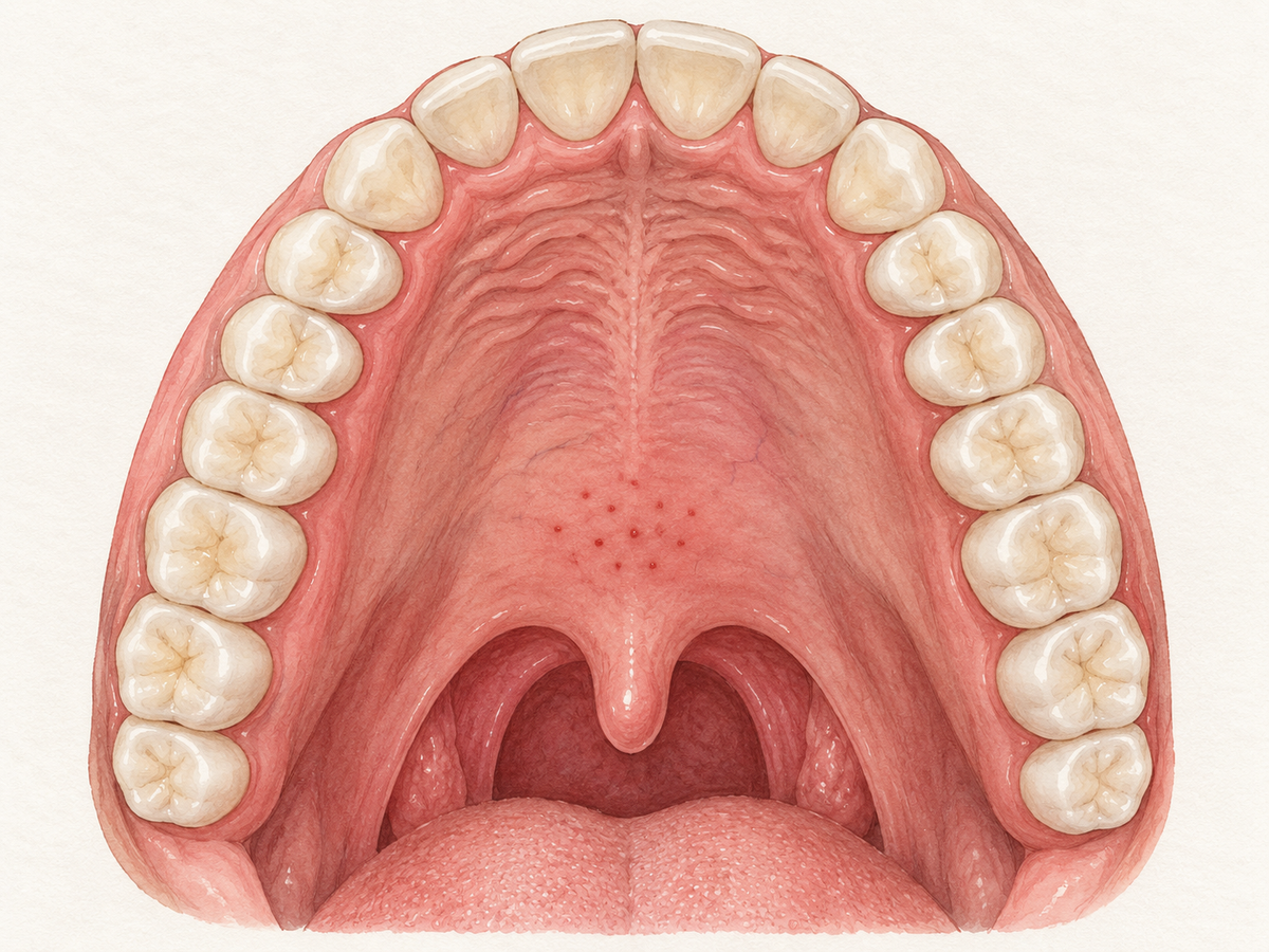
Red spots on the roof of your mouth are usually not serious. The most common causes are everyday irritation or minor injury, such as hot food, a scratchy chip, dentures or braces, or suction from coughing or oral sex. Most of these spots heal on their own within a few days to two weeks. But a spot that persists, grows, bleeds, or comes with fever, severe pain, or trouble swallowing or breathing should be checked by a dentist or doctor.
Use a simple rule: if you can link a flat, small, painless spot to a clear irritation or recent injury, watch it for a week or two. If it does not improve within two weeks, or if you feel unwell, get it checked.
Start With the Spot Pattern
The way a spot looks helps narrow the cause. A flat pinpoint dot often behaves differently from a raised bump, a larger flat red area, or a painful open sore.
- Petechiae are flat pinpoint red spots. They happen when tiny blood vessels under the mouth lining leak a little blood.
- Papules are raised bumps.
- Patches are larger flat red areas.
- Oral ulcers are painful open sores, sometimes with a white or yellow center.
- Erythema means general redness without a clear sore or bump.
The pattern alone is not enough. A red spot can come from harmless irritation, infection, an immune reaction, or rarely a precancerous change, so clinicians also look at the story behind it. A review of red oral lesions explains that the spot pattern plus your history helps narrow the possibilities.
Along with the pattern, note how long it has been there, what may have triggered it, and whether it bleeds or comes back. That is the information a dentist or doctor will ask for. Flat pinpoint spots called petechiae can appear in many conditions, so duration and triggers are especially useful.
Everyday Irritation and Injury
Most red spots on the palate come from simple irritation or minor injury.
Common causes include:
- Burns from hot pizza, coffee, or other hot foods and drinks.
- Friction from dentures, braces, hard foods, or vigorous brushing.
- Suction from coughing, vomiting, forceful sucking on straws, or oral sex.
Suction can pull on small blood vessels and create flat pinpoint spots, especially on the soft palate. A case review of diffuse oral petechiae notes that repeated negative pressure from forceful sucking or thrusting can cause these harmless pinpoint spots. In another report, palatal spots related to oral sex healed on their own within two weeks without treatment.
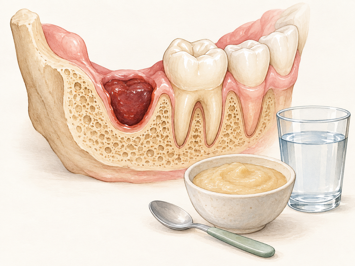
Denture-related spots or sores usually improve after the denture is adjusted or left out for a while. Traumatic denture ulcers often heal within about a week after the cause is removed. The key step is to stop the trigger and watch for improvement.
Infections That Target the Palate
Some infections can cause red spots or sores on the roof of the mouth, often with other symptoms like fever, sore throat, or tiredness.
Viral causes include:
- Herpes simplex, the cold sore virus, which can also cause mouth sores.
- Hand-foot-and-mouth disease and herpangina, which commonly cause mouth ulcers in children.
- Strep throat, a bacterial infection that can produce tiny red spots on the palate along with a sore throat and fever.
- Mononucleosis or mono, caused by the Epstein-Barr virus, which may cause palatal petechiae with severe sore throat and fatigue.
A review of childhood oral infections describes these viral illnesses as common causes of oral sores and spots. For strep throat, clinical guidelines list palatal petechiae as one possible sign, but also note that symptoms alone cannot reliably separate strep from a viral throat infection. A review of pediatric pharyngitis adds that palatal petechiae may point toward strep or Epstein-Barr infection.
Fungal infections can also appear as red areas. The red form of oral thrush, sometimes called erythematous candidiasis, is often seen under a denture. It can be worse with dry mouth, recent antibiotics, or poor oral hygiene. Denture-related changes are common, and reviews of red oral lesions describe denture stomatitis as a red, usually painless change limited to the denture-bearing area.
If spots are spreading, or if they come with fever, sore throat, or feeling unwell, see a clinician for an exam and possible testing.
Oral and Systemic Conditions to Consider
Less commonly, red spots in the mouth can be part of a body-wide issue.
Immune-related conditions can affect the mouth lining:
- Oral lichen planus is a chronic inflammatory condition that can cause red, tender, or white patches.
- Pemphigus and pemphigoid are autoimmune blistering diseases that may cause painful erosions or ulcers on the gums, cheeks, or palate.
A review of oral lesions in autoimmune blistering diseases explains that these conditions can look similar, so diagnosis often requires a biopsy and special tests.
Blood or platelet problems may show up as pinpoints, purplish spots, easy bruising, or gum bleeding. A review of oral signs in blood disorders notes that platelet problems can cause facial or oral petechiae and bleeding. Nutritional deficiencies, such as low vitamin C, iron, or B vitamins, can also make the mouth lining red, sore, or prone to bleeding. Nutritional deficiencies and oral signs are worth considering when oral spots appear along with fatigue or a limited diet.
Medication or allergic reactions can also contribute. Blood thinners and certain herbal supplements may make pinpoint spots more likely, according to a review of diffuse oral petechiae.
These causes are less common, but they become more likely when spots recur or come with skin, eye, or genital symptoms, fatigue, easy bruising, or a recent medication change. When nothing local explains the spots, blood work or a specialist referral may be needed.
Red Flags That Need Prompt Care
Some signs should not wait for home remedies.
- A single red patch or lump that does not heal within two weeks.
- Bleeding from the spot.
- Swelling that is getting larger.
- Severe pain or fever.
- Difficulty swallowing or breathing.
- Spots linked to smoking, chewing tobacco, or heavy alcohol use.
A velvety or granular red patch that persists can be a concern because some red patches, called erythroplakia, are more likely to contain precancerous or cancerous cells. A review of red oral lesions reports that a high proportion of true erythroplakia cases show concerning changes on biopsy. Referral guidance also recommends urgent assessment for a red or red-and-white patch in the mouth. Tobacco and alcohol exposure can contribute to these red or white patches, so mention those habits to your clinician.
These red flags could point to a spreading infection, a blood disorder, or oral cancer. Contact a dentist, physician, or urgent care depending on severity.
What to Do While You Wait
If there are no red flags, you can take simple steps at home.
- Avoid hot, spicy, and acidic foods and drinks that can sting or irritate.
- If dentures or other appliances are rubbing, stop wearing them as much as possible until they can be adjusted.
- Rinse gently with a plain salt-water rinse to keep the area clean and soothe it.
- Take photos once a day to track changes in size, color, or number.
- Do not pick at spots or apply unknown remedies, which can make irritation worse.
If the spots worsen, new symptoms appear, or they are still present after two weeks, book a dental or medical exam.
How a Clinician Narrows It Down
The work-up usually starts with a conversation and a mouth exam.
Your clinician will ask about how long the spots have been present, recent foods, injuries, illnesses, medications, smoking, and alcohol use. They will look at the size, shape, color, and location of the spots.
If needed, they may use:
- A swab to test for viral, bacterial, or fungal infection.
- Blood tests to check for blood disorders, platelet problems, or nutritional deficiencies.
- An allergy review to look for medication or contact reactions.
- A biopsy, or small tissue sample, when a persistent patch could be precancerous or autoimmune.
A review of oral petechiae diagnosis describes this kind of step-by-step assessment: oral exam, history, recent activity, and blood tests when needed. Biopsy is used when a red patch cannot be explained by more common causes or when cancer is suspected.
Most causes are diagnosed without invasive testing. If a spot lasts, call a dentist. If you also have fever, severe pain, or other body-wide symptoms, contact a doctor.
This article has been reviewed by an oral health professional for accuracy. It is intended to provide general educational information and should not be used as a substitute for personalized medical advice, diagnosis, or treatment. If you have questions or concerns about your oral health, please consult a dentist, physician, or other qualified healthcare provider.